
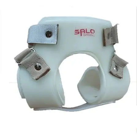
CDH Splint
Features
- Maintains Hip in Abduction for Concentric Femoral Head Reduction & Acetabular Development
- Lightweight, Washable & Easily Adjusted for Maternal & Carer Compliance
- First-Line Conservative Alternative to Spica Casting & Surgical Intervention
- Clinically Effective Across Graf Types IIa, IIb, IIc & Unstable Hip Presentations
- Open Diaper Access Design for Practical Neonatal Hygiene Management
- Promotes Natural Hip Movement While Maintaining Therapeutic Abduction Position
- Suitable for Full-Time & Nocturnal Wearing Schedules Across Severity Presentations
CDH Splint Paediatric Hip Abduction Orthosis for Congenital Dislocation of the Hip, Neonatal Hip Instability & Early Developmental Dysplasia Management
The CDH Splint is a purpose-designed paediatric hip abduction orthosis developed specifically for the conservative management of Congenital Dislocation of the Hip (CDH) now more comprehensively termed Developmental Dysplasia of the Hip (DDH) the most common congenital musculoskeletal condition in newborns, encompassing a spectrum of hip pathology ranging from mild acetabular dysplasia without femoral head displacement through the frankly dislocated hip in which the femoral head is entirely outside the acetabular socket. Congenital dislocation of the hip remains a significant problem in terms of diagnosis and treatment the imperative is early diagnosis since most hips will respond to simple abduction splintage, while a later diagnosis is synonymous with surgical intervention either in the form of open or closed reduction. The CDH splint operationalises this clinical principle providing the abduction positioning that enables the biological self-correction of the dysplastic hip during the most therapeutically responsive period of the infant's skeletal development, in the most practically manageable, parent-friendly, and developmentally appropriate orthotic format available.
Understanding CDH/DDH: Prevalence, Aetiology & the Critical Importance of Early Treatment
The incidence of neonatal hip instability is around 15–20 per 1000 live births, but that of established dislocation 1–2 per 1000 live births in unscreened cohorts. This epidemiological distinction between hip instability and established dislocation reflects the important biological reality that the majority of neonatal hip instability presentations resolve spontaneously during the first weeks of life as the neonatal ligamentous laxity driven by the maternal relaxin hormone present in the fetal circulation progressively resolves and the periarticular capsular and muscular restraints mature. However, most of these resolve without specific treatment, with only ten percent going on to eventually dislocate, and a further ten percent showing signs of dysplasia.
The clinical challenge is identifying which unstable hips will self-resolve and which require active orthotic intervention a challenge that ultrasound screening based on Graf's classification system is now widely used to address. Given the existing scientific evidence and changes in newborn DDH screening practices, universal ultrasound screening seems to be the best option for preventing late-detected cases and can be recommended as a favourable prevention strategy. When ultrasound confirms persisting instability or dysplasia beyond the expected self-resolution window, active CDH splint treatment is initiated the clinical decision point at which the orthotist and paediatric orthopaedic surgeon must prescribe the correct abduction orthosis to achieve the concentric femoral head reduction that drives acetabular development.
A total of 20% of children with DDH will not have stood up or walked by 18 months compared with just 5% of the normal population a statistic that powerfully illustrates the functional developmental impact of inadequately managed hip dysplasia and the clinical importance of effective early orthotic intervention in preventing the mobility limitation, gait abnormality, and early hip osteoarthritis that are the long-term consequences of untreated or undertreated CDH.
The Therapeutic Mechanism: Why Hip Abduction Corrects CDH
The therapeutic mechanism of the CDH splint and the biological rationale for the hip abduction position it maintains is rooted in the pressure-mediated remodelling properties of the immature acetabular cartilage. The neonatal acetabulum is composed predominantly of cartilage rather than bone a biological state that makes it both susceptible to deformation under abnormal loading and highly responsive to corrective mechanical stimulation during this critical period of skeletal plasticity.
When the CDH splint positions the hip in abduction and flexion, the femoral head is placed in direct, concentric contact with the acetabular cartilage at its most central, deepest point. This sustained contact generates a gentle, evenly distributed compressive stimulus across the acetabular cartilage surface a stimulus that, in accordance with the biological principle that cartilage and bone remodel in response to mechanical loading patterns, drives the progressive deepening and lateral coverage improvement of the acetabular socket. The acetabulum literally remodels around the femoral head that is consistently maintained within it, developing the socket depth and coverage angle that constitutes normal hip joint architecture and that is the biological goal of all CDH conservative management.
The Design Philosophy: Compliance Above All Other Considerations
A lightweight, washable, and easily adjusted splint for the congenitally dislocated hip, designed to improve maternal compliance, is described. This deceptively simple design brief encapsulates the most critical clinical insight in CDH splint design that the therapeutic effectiveness of any hip abduction orthosis is entirely dependent on the consistency and duration of its application by the infant's parents and carers, and that every design feature of the CDH splint must therefore be evaluated primarily through the lens of its contribution to parental compliance.
A splint that is difficult to apply, impossible to keep clean, uncomfortable for the infant, or disruptive to normal feeding and care routines will be used inconsistently, modified incorrectly, or abandoned prematurely outcomes that are clinically equivalent to no treatment at all, and that allow the hip dysplasia to progress during the precise developmental window when biological correction was achievable. The CDH splint's combination of lightweight construction, machine-washable materials, straightforward adjustment mechanisms, and open diaper access design directly addresses each of the practical barriers to compliance that are most commonly cited by families managing CDH splint treatment across the weeks and months required for effective hip reduction and acetabular development.
Avoiding Avascular Necrosis: The Critical Safety Consideration in CDH Splinting
Early splinting in abduction, however, may occasionally result in avascular necrosis of the femoral head. Avascular necrosis (AVN) of the femoral head the ischaemic death of the cartilaginous and bony tissue of the femoral head due to interruption of its blood supply is the most serious complication of CDH orthotic treatment, and the primary design consideration that distinguishes modern CDH splints from the rigid abduction frames of historical practice. The blood supply to the neonatal femoral head is particularly vulnerable to compression from excessive or forcibly applied abduction a clinical reality that drove the progressive evolution of CDH splint design away from rigid, fixed-position immobilisation devices towards the dynamic, movement-permitting designs that characterise contemporary clinical practice.
The design comes as close as possible to a dynamic positioning orthosis differing from earlier abduction splints which completely immobilised the hips and therefore caused a high rate of avascular necrosis the CDH splint achieves its corrective abduction positioning while permitting sufficient freedom of lower limb movement to maintain the dynamic loading and unloading of the femoral head that supports its normal vascular perfusion during treatment. This dynamic approach maintaining the therapeutic abduction angle while allowing the natural kicking and movement of the infant represents the clinical consensus refinement of CDH splinting methodology across decades of clinical observation and outcome data.
Graf Classification & Treatment Prescription: Matching Splint Use to Diagnostic Category
The Graf ultrasound classification system which assigns hip joints to types based on the alpha angle (measure of acetabular bony coverage) and beta angle (measure of cartilaginous coverage) observed on standardised coronal ultrasound images provides the diagnostic framework within which CDH splint prescription is made. Graf type I hips are normal and require no treatment. Graf type IIa hips in infants under three months represent physiological immaturity and are managed with watchful waiting and repeat ultrasound. Graf types IIb and IIc represent established dysplasia requiring active orthotic treatment. Graf type III hips are dislocated and require active reduction and maintained abduction. Graf type IV hips are severely dislocated and typically require more intensive intervention.
The CDH splint is clinically indicated across the Graf types IIb through III range presentations that encompass the vast majority of clinically significant hip dysplasia cases encountered in neonatal hip screening programmes. The abduction brace allowed reduction in 85% of patients across these Graf types confirming the CDH splint's capacity to achieve and maintain concentric femoral head reduction in the large majority of dysplastic hips that fall within its indication range, representing an evidence-validated first-line treatment approach that prevents the progression to surgical intervention in the great majority of diagnosed cases when initiated promptly.
Wearing Schedule, Clinical Monitoring & Treatment Duration
The CDH splint wearing schedule is determined by the treating orthopaedic surgeon and orthotist based on the severity of the dysplasia, the infant's age at diagnosis, the degree of hip reduction achieved at initial application, and the ultrasound response to treatment at each clinical review. Full-time wearing 23 to 24 hours per day is typically prescribed for frank dislocations and significant dysplasia requiring active maintained reduction, with the splint removed only for bathing and diaper changes. After initial reduction, splints were worn part-time, at night, for two months the progressive reduction in wearing time as the hip responds to treatment and the acetabular development improves reflects the staged approach to CDH treatment discontinuation that minimises the risk of dysplasia recurrence during the early consolidation phase following active treatment cessation.
Clinical and ultrasound review at regular intervals typically every four to six weeks during active treatment allows the treating team to assess the acetabular development trajectory, document the improving alpha angle, confirm maintained femoral head centring, and adjust the wearing schedule and abduction angle prescription in response to the individual infant's treatment response. Skin inspection at each review identifies any pressure-related concerns at the splint contact zones particularly at the femoral condyles, greater trochanter, and perineal region before they progress to skin breakdown.
Practical Guidance for Families: Application, Care & Daily Management
Families managing CDH splint treatment require comprehensive education at each clinical contact regarding correct splint application technique, strap tension adjustment, diaper change management within the splint, bathing with the splint removed, monitoring for signs of skin irritation or pressure, recognising when the splint requires adjustment due to growth, and understanding the clinical importance of consistent wearing schedule compliance for treatment success. The CDH splint's washable materials allow routine cleaning with mild soap and water maintaining the hygienic standards required for a device worn in close proximity to the perineal region of a nappy-wearing infant across 24-hour daily use periods. Growth-related size adjustments are expected at intervals of four to eight weeks during the rapid growth of early infancy, and families should be advised to contact their orthotist promptly if the splint appears loose, shifts position during use, or creates any marks on the infant's skin that persist beyond 20 minutes after removal.
Indicated For:
Congenital dislocation of the hip (CDH), developmental dysplasia of the hip (DDH) across Graf types IIb, IIc, III, and selected type D presentations, neonatal hip instability detected on Barlow and Ortolani testing with ultrasound confirmation, acetabular dysplasia in infants and young children up to 18 months of age amenable to conservative abduction splint treatment, post-closed reduction hip maintenance in appropriate presentations, unilateral and bilateral hip dysplasia, hip dysplasia in infants with risk factors including breech presentation, oligohydramnios, first-degree family history, and talipes, and any neonatal or early infant hip presentation requiring maintained passive hip abduction positioning to achieve and sustain concentric femoral head reduction and promote acetabular socket development during the biologically responsive period of early skeletal growth.
Add your review
Your email address will not be published. Required fields are marked *
Please login to write review!
Looks like there are no reviews yet.

M. A. Medical Supports