
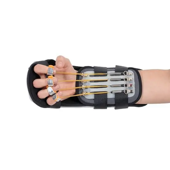
Kleinert Splint
Features
- Dynamic Rubber Band Traction for Passive Flexion & Active Extension Protocol
- Wrist in 45° Flexion & MP Joints in 10–20° Flexion for Optimal Repair Protection
- Palmar Pulley System for Enhanced PIP & DIP Passive Flexion
- Active Extension Protocol Inhibits Flexor Muscle Contraction for Safe Tendon Loading
- Clinically Proven Outcomes Across Flexor Tendon Repair Zones I–V
- PIP Flexion Contracture Prevention Through Night-Time Strap Protocol
- Three-Stage Progressive Protocol: Passive Motion to Full Active Rehabilitation
Kleinert Splint Dynamic Dorsal Blocking Orthosis for Post-Operative Flexor Tendon Repair, Controlled Passive Mobilisation & Adhesion Prevention
The Kleinert Splint formally known as the Kleinert Dynamic Dorsal Blocking Splint is the most historically significant and clinically foundational dynamic orthosis in post-operative flexor tendon rehabilitation. Introduced by Harold Kleinert in the 1950s and progressively refined through decades of clinical application and research, the Kleinert splint and its associated rubber band passive flexion protocol represented a paradigm-shifting departure from the complete immobilisation that had previously been the standard of care following flexor tendon repair establishing the principle of controlled early passive mobilisation as the biological and biomechanical foundation upon which all subsequent flexor tendon rehabilitation philosophy has been built. The emergence of the Kleinert regimen was a breakthrough, promoting early movement of the affected digits by the aid of a rubber band, with the efficiency of the Kleinert protocol subsequently verified by numerous clinical studies over more than half a century of hand surgery practice.
The Clinical Problem: Why Flexor Tendon Repair Demands Specialised Orthotic Management
The flexor tendons of the hand are among the most biomechanically demanding structures in the musculoskeletal system subjected to exceptionally high tensile loads during grip and pinch, enclosed within a precisely engineered fibro-osseous sheath system that must allow near-frictionless gliding across the full range of digital motion, and repaired in a biological environment where the competing demands of healing strength and mobility prevention place the treating hand surgeon and therapist in a perpetual therapeutic tension. Flexor tendon injuries without surgical repair do not heal the disrupted tendon ends retract, the sheath collapses, and the functional deficit is permanent. However, surgical repair itself creates a new set of biological challenges: the suture site, surrounded by the inflammatory exudate of the healing response, is the epicentre of the adhesion formation process that threatens to bond the repaired tendon to its surrounding sheath and eliminate the tendon gliding that is the prerequisite for active digital flexion.
The Kleinert splint directly addresses both of these competing biological imperatives protecting the repair site from the tensile overload that would rupture the suture while simultaneously enabling the controlled tendon gliding that prevents the adhesion formation that would render the repair functionally useless despite structural integrity.
The Biomechanical Mechanism: Active Extension Against Rubber Band Resistance
The therapeutic mechanism of the Kleinert splint is both elegantly simple and profoundly biomechanically sophisticated. The rubber band, attached from the fingernail to an anchor point at the wrist or volar forearm, generates a continuous, low-load passive flexion force on the involved digit maintaining the finger in a passively flexed position at rest. When the patient performs their hourly exercise sessions, they actively extend the finger against the rubber band's resistance until they reach the extension limit imposed by the dorsal blocking splint. As they relax after each active extension, the rubber band passively returns the finger to the flexed position.
This active extension phase generates the critical tendon excursion that prevents adhesion formation the differential gliding between the repaired tendon and its surrounding sheath structures that disrupts the early, immature adhesion bonds before they organise into the mature, mechanically resistant fibrotic adhesions that permanently restrict tendon gliding. Crucially, this tendon excursion is achieved during the active extension phase when the flexor tendon is being paid out proximally not during active flexion, which would generate active flexor muscle contraction and potentially catastrophic tensile stress at the suture line. The passive flexion return phase provided by the rubber band requires no active flexor muscle contraction, protecting the repair from the tensile forces of voluntary flexor activation throughout the entire post-operative protocol period.
The Dorsal Blocking Splint: Defining the Protected Range
The dorsal blocking component of the Kleinert splint is not merely a structural housing for the rubber band system it is an active and essential biomechanical element that defines the range within which the controlled passive mobilisation protocol operates and determines the protective limits that prevent repair rupture. The wrist flexion position of 45 degrees is the most important single positioning parameter of the Kleinert splint: by placing the wrist in flexion, the effective working length of the flexor tendon pathway from muscle origin to digital insertion is shortened, reducing the tensile stress generated at the suture line during each active extension exercise to a level well below the mechanical strength of the healing repair at each post-operative timepoint.
The MCP joint position at 10–20 degrees of flexion serves a complementary protective function maintaining the collateral ligaments of the MCP joints at near-maximum length to prevent the MCP extension contracture that rapidly develops when the MCP joints are held in extension during the period of post-operative swelling and reduced mobility. The IP joints are held in extension by the dorsal blocking structure between exercise sessions, maintaining the volar plate at adequate length and preventing the PIP flexion contracture that is the Kleinert protocol's most prevalent complication.
The Palmar Pulley Modification: Addressing the PIP & DIP Flexion Deficit
The original Kleinert technique with the rubber band anchored directly at the wrist was recognised early in its clinical application to produce incomplete passive flexion at the PIP and DIP joints, as the proximal rubber band anchor created a moment arm that preferentially flexed the MCP joint while delivering insufficient flexion force distally. The palmar pulley modification directly addresses this limitation by rerouting the rubber band through a transversely positioned pulley at the level of the distal palmar crease, changing the direction of the traction force and dramatically increasing the passive flexion delivered at the PIP and DIP joints. The modified Kleinert splint increases passive flexion at the PIP and DIP joints, achieving more complete composite digital flexion the position in which the flexor digitorum profundus achieves maximum excursion at the repair site making the modified Kleinert with palmar pulley the preferred configuration for the majority of hand centres currently using passive motion protocols.
Three-Stage Progressive Rehabilitation: From Passive Motion to Full Function
The Kleinert protocol follows a precisely structured three-stage progression that advances the mechanical demands placed on the healing tendon in strict synchrony with the biological timeline of tendon repair consolidation. The early stage commencing at three to five days post-operatively and continuing for the first three weeks establishes the rubber band passive flexion/active extension exercise routine that is the foundation of the protocol, with the patient performing ten active extension repetitions within the dorsal block every hour throughout the waking day. The recommended frequency of rubber band-assisted passive flexion–active extension exercises was six to eight times a day during the initial weeks, ensuring sufficient tendon excursion frequency to prevent adhesion organisation while allowing adequate repair site rest between exercise bouts.
The intermediate stage at three to four weeks transitions from the full dorsal blocking splint to a wrist band assembly that maintains the rubber band traction attachment without the full dorsal blocking extension, enabling progressive wrist and hand mobilisation while continuing to protect the repair site from excessive tensile loading during the phase of early collagen deposition and remodelling. The advanced stage from four to six weeks advances to active motion, place-and-hold exercises, and progressive removal of the splinting system as the tendon approaches sufficient mechanical strength to tolerate active loading without rupture risk.
Complications, Monitoring & Clinical Management
The PIP flexion contracture the most prevalent and functionally significant complication of the standard Kleinert rubber band traction protocol requires proactive clinical monitoring and early intervention at every post-operative review. When evidence of developing PIP flexion contracture is detected, the standard management response is to remove the rubber band traction at night and strap the IP joints into extension against the dorsal block, allowing the continuous overnight passive extension to counteract the contractile forces that the daytime rubber band traction creates. The clinical team must balance the conflicting demands of preventing adhesion formation through adequate passive flexion during the day and preventing PIP contracture through adequate IP extension at night a balance that requires regular, skilled clinical assessment and personalised protocol modification.
Patients are assessed at weekly intervals during the first post-operative month, with tendon integrity confirmed by evaluating passive and active motion, documenting total active motion (TAM) and tip-to-palm distance, and monitoring for the signs of repair rupture the sudden loss of passive flexion resistance and ability to extend the digit fully or developing adhesion formation the increasing discrepancy between passive and active flexion range that indicates tendon adherence to surrounding structures.
Comparative Evidence: Kleinert vs Alternative Protocols
The clinical evidence comparing the Kleinert passive motion protocol to alternative flexor tendon rehabilitation approaches including the Duran controlled passive motion protocol and contemporary early active motion protocols consistently demonstrates that the Kleinert protocol achieves clinically significant tendon excursion and functional outcomes, with early passive motion protocols including both Duran and Kleinert types resulting in a 4% rupture rate and 9% decreased range of motion across 1,598 tendon repairs. Early active motion protocols demonstrate slightly higher rupture rates but lower rates of restricted range of motion, reflecting the fundamental biomechanical trade-off between repair protection and adhesion prevention that defines the flexor tendon rehabilitation challenge. The Kleinert protocol's established safety profile and well-documented outcome data make it the reference standard against which all newer protocols are compared in contemporary hand therapy research.
Indicated For:
Primary flexor tendon repair in Zones I, II, III, IV, and V post-operatively, secondary flexor tendon repair and reconstruction, flexor tendon repair in the thumb, post-operative flexor tenolysis (where gentle early motion is required to maintain the adhesion-free gliding achieved surgically), paediatric flexor tendon repair where a less demanding early active protocol is clinically preferred, and any post-operative flexor tendon management scenario where a clinician-prescribed passive flexion/active extension early mobilisation protocol is the selected rehabilitation approach.
Add your review
Your email address will not be published. Required fields are marked *
Please login to write review!
Looks like there are no reviews yet.

M. A. Medical Supports